
Imagine the remarkable ability to visualize the intricate inner workings of our organs and tissues in the human body. This is made possible by emission tomography scanners, advanced medical imaging devices that provide detailed images of the body’s internal structures.
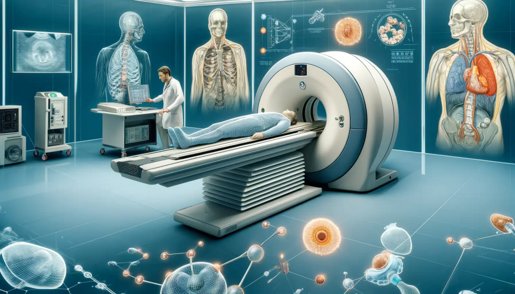
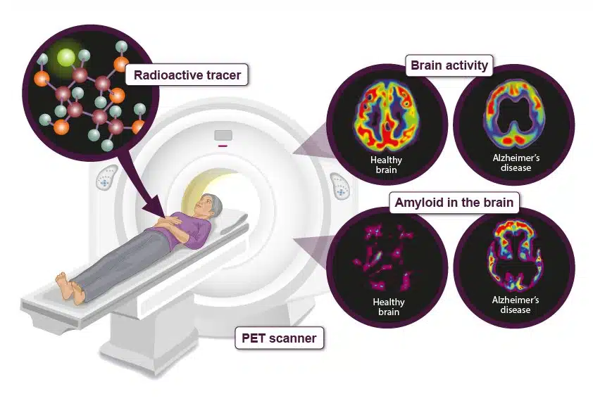
The functioning principle of these scanners involves the detection and measurement of energy emitted by radioactive substances, known as radiotracers, which are administered into the body. Similar to miniature beacons, these radiotracers emit energy that can be detected by the scanner, enabling the creation of precise mappings of internal structures.
Emission tomography scanners come in various types, each possessing distinct capabilities. Positron emission tomography (PET) scanners utilize radiotracers emitting positrons, minuscule particles that interact with electrons to generate gamma rays. On the other hand, single-photon emission computed tomography (SPECT) scanners employ radiotracers directly emitting gamma rays.
As the radiotracers undergo decay, they emit gamma rays that the scanner detects. Utilizing this information, the scanner generates three-dimensional images of internal organs and tissues. These images serve in diagnosing and monitoring a wide range of conditions, including cancer, heart disease, and neurological disorders.
Emission tomography scanners find extensive usage in clinical practice, recognized for their safety and effectiveness. However, it is essential to consider the benefits and risks associated with ionizing radiation exposure before proceeding with the procedure.
In conclusion, emission tomography scanners represent powerful tools that offer unparalleled insights into the human body’s internal workings. By detecting and measuring energy emitted by radioactive substances, these scanners facilitate the creation of detailed images, aiding doctors in diagnosing and monitoring various medical conditions. Although exposure to ionizing radiation is involved, the benefits of these scanners often outweigh the associated risks, solidifying their indispensability in modern medicine.
Top 10 fun facts about emission tomography scanners
- The pioneers of the PET scanner were a team of researchers at Washington University in St. Louis, who successfully developed the first PET scanner during the 1970s.
- Emission tomography scanners utilize radiotracers that are typically created by combining a radioactive atom with a specially designed molecule, enabling them to target specific organs or tissues in the body.
- PET scanners possess the capability to measure metabolic activity within the body, which aids doctors in diagnosing and monitoring various conditions, including cancer and neurological disorders.
- SPECT scanners are valuable tools for studying blood flow in the brain and detecting abnormalities in the heart.
- The radiation exposure associated with a typical PET scan is relatively low, equivalent to approximately three years’ worth of natural background radiation.
- In neuroscience research, PET scanners are utilized to investigate brain activity in response to different stimuli, such as visual or auditory cues.
- SPECT scanners find frequent usage in nuclear medicine for the diagnosis and monitoring of bone diseases, including osteoporosis and bone cancer.
- Sports medicine has benefited from the use of PET scanners, which enable the study of metabolic activity in muscles during exercise.
- PET scanners have played a role in psychology research, providing insights into the neural correlates of emotions, motivation, and other cognitive processes.
- The advent of emission tomography scanners has revolutionized the field of medical imaging, profoundly impacting the diagnosis and treatment of a wide range of diseases and conditions.